Novel Technique Improves Brain-State Detection
Mathematics-based approach makes functional near-infrared spectroscopy more reliable
• New method boosts precision of a non-invasive brain-state classification with functional near-infrared spectroscopy (fNIRS).
• Achieved high accuracy, outperforming traditional approaches.
• Advances fNIRS as a reliable, accessible tool for brain measurements.
• Method has potential as a diagnostic tool for vegetative states.
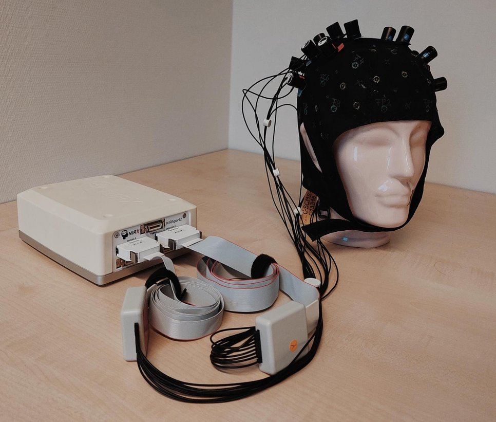
Researchers have developed a new method that greatly improves the accuracy of brain-state classification with functional near-infrared spectroscopy (fNIRS). The brain-imaging technique fNIRS allows researchers to measure neural activity: Active brain cells need more oxygen, so variations in blood flow and oxygen saturation indicate which brain regions are at work. fNIRS detects these changes safely and without invasive procedures.
Despite its many advantages in clinical settings – it is portable, cost-effective, and reliable even if the patient moves –, the methods for analyzing fNIRS data are less advanced compared to other brain-imaging techniques.
Harnessing the dual nature of fNIRS signals

An international research team has now developed a method for classifying brain states with fNIRS which is specifically tailored to the unique properties of the fNIRS signal and achieves therefore more accurate results. In contrast to other brain imaging methods, fNIRS measures both the oxygenated and the deoxygenated blood. “These two signals naturally show opposite patterns, but that doesn’t mean they are redundant,” explains Tim Näher, first author of the study, who works at the Max Planck Institute for Biological Cybernetics in Tübingen, Germany. “Instead, they give us complementary insights into brain activity.” Näher and his coauthors exploited this unique dual nature of the fNIRS signal by applying advanced mathematical tools from a field called Riemannian geometry.
To test their new method, the team asked healthy participants to perform eight different mental tasks such as imagining playing tennis, internally singing, or rotating an object in their mind. With the new computational framework, the researchers were able to classify very accurately which of the tasks a participant was executing, significantly surpassing the precision of traditional methods.
New approach may help reveal signs of consciousness in patients
The results could play an important role in improving the diagnosis of disorders of consciousness. These conditions are notoriously difficult to assess: Patients often have little to no ability to move or communicate, making it challenging to determine whether they retain some level of wakefulness and awareness. However, an accurate diagnosis is essential for effective treatment and reliable prognosis.
This is why Näher collaborated with Lisa Bastian of the University of Tübingen on a second study, conducted at Bettina Sorger’s lab at Maastricht University, which developed a novel fNIRS paradigm to assess whether a non-responsive patient is still conscious. To this end, healthy participants were asked either to carry out a mental task—simulating a state of awareness—or to remain consistently inactive. The novel fNIRS paradigm combined with the new data-analysis approach based on Riemannian geometry proved remarkably accurate in distinguishing between responsive and unresponsive brain states: It correctly identified responsiveness every time and accurately recognized unresponsiveness in nine out of ten cases.
“So far, we provided a proof of concept that the new fNIRS framework can serve as a fast, objective, and accessible tool to support more reliable diagnoses and improve treatment decisions for disorders of consciousness,” Näher says. “The next logical step would be to test our method in real patients.”
